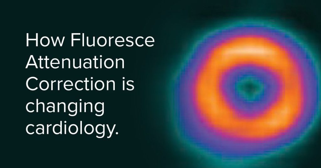
Attenuation correction is a mechanism that removes soft tissue artifacts from nuclear images. The goal is to reduce the impact of attenuation in order to provide images that are more uniform and allow for higher reading confidence.
Historically, Positron Emission Tomography (PET) imaging was the only way to achieve attenuation correction and offered features that were, at one time, a distinct advantage over SPECT cameras.
However, with the advancements in solid-state technology and attenuation correction methods, specifically Fluoresce Attenuation Correction (FAC), SPECT can now deliver higher quality, PET-like image resolution, greater diagnostic accuracy, and higher reading confidence than ever before.
Fluoresce attenuation correction
Attenuation correction can provide valuable diagnostic data in cardiac imaging interpretation. Fluoresce Attenuation Correction is a method that utilizes a fluorescence x-ray, which significantly contributes a better, cleaner image, that allows for a lower dose and less radiation exposure to the patient. It’s a unique combination of hardware and software technology that delivers superior image quality with the lowest possible radiation burden.
The combination of FAC and SPECT
By leveraging FAC, SPECT is able to identify false artifacts, correct, and capture the accurate distribution of the imaging agent, thus allowing for higher reading confidence and diagnostic accuracy.
All in all, it can reduce the number of false positives and inconclusive studies that could potentially lead to unnecessary tests or invasive procedures like cardiac catheterizations.
Ultimately, SPECT with FAC offers PET-like image quality and is much more cost effective in terms of hardware and ongoing consumables. It also does not necessitate the room shielding and logistics that PET requires. In the end, Fluorescence Attenuation Correction delivers a significantly higher image quality for a substantially lower cost.
FAC and the Digirad X-ACT+
Fluorescence attenuation correction is available in conjunction with the Digirad X-ACT+ camera. The X-ACT+ is the only SPECT/FAC MPI system that features a combination of solid state detectors, rapid imaging detector geometry, low dose fluorescence x-ray attenuation correction, advanced 3D-OSEM reconstruction techniques, and TruACQ Count Based Imaging™. It offers high definition, high efficiency, unparalleled clinical accuracy—all while lowering the patient’s radiation dose. If you’d like more information on the X-ACT+ camera, download a brochure or request a consultation here.