If you diagnose or treat patients with liver cancer, then you’re probably familiar with the procedure for Yttrium-90 (Y90) studies.
In short, the patient must go through a mapping process to ensure that the medical team gets the best possible information from which to treat them. This involves examining the vasculature of the tumor and the interventional radiologist may place coils or perform embolizations; a deliberate blockage of the vasculature to limit the blood flow to and from the intended area of treatment.
With embolization complete, the patient is administered a radioactive particulate (99Tc-MAA) that is similar in size to Y90 . They are then sent for imaging using a gamma camera to assess the liver and lungs. This determines whether the patient is ready for the Y90 treatment or whether they require further embolization to to ensure that the Y90 will be directed to the intended location within the tumor.
It’s a minimally invasive process, but it still can be hard on the ill patient and somewhat inefficient for the medical facility. The type of camera you use matters, as does the modality of the camera.
Inefficiencies with Y90 procedures
Traditionally, as the patient undergoes the described process, they will be transported from the procedure room to the nuclear medicine suite for imaging. The aim of the first set of images is to check that embolization has been successful – meaning that 80% or more of the 99Tc-MAA is localized within the liver and does not shunt to the lungs.
If the finding is that 20% or more of the particulate dose does go to the lungs, then the decision will often be to send the patient back for further embolization. This is done to ensure that when the Y90 is administered, it reaches the intended location within the liver.
Here’s where that traditional approach can be inefficient:
- You’re transporting the patient from one area of the hospital to another for imaging. This involves keeping them sedated as well as an anesthesia team to transport with them.
- The camera, an expensive investment for the hospital is kept open, waiting for the patient having the mapping procedure.
- The process may have to be repeated again if the target area is not being reached. This means that the camera is kept open for an even longer period and is unable to be used for other patients.
These points can mean that the hospital staff are unable to use their time and resources as efficiently as they could. It is usually the aim to be able to make the best use of time and equipment, so that patient throughput is maximized.
From the patient’s perspective
The patient is kept sedated throughout this process, however it is within their best interests to minimize time under anesthesia. The “transport then repeat” scenario can effectively double or more their time under sedation. It also means that more of their day is taken up undergoing the procedure.
You also should consider the risk to the patient whenever they are required to be transported in the hospital. It is well-documented that there are many risks to patients during transportation and that an approach to minimize the need for transportation is safer. For example, they risk exposure to infectious diseases, dislodgement of equipment and associated injuries.
The bottom line is that quicker studies with less time under sedation and removing the need for transport are a better experience for the patient too.
Mobile imaging: A solution for Y90 therapy
There is a more efficient solution to streamlining Y90 imaging; a mobile gamma camera can be brought into the treatment area. This removes the need to transport the patient and allows any fixed cameras to remain in use throughout the procedure. It is also a good measure to improve patient safety.
The type of camera used is also important. An issue with traditional gamma cameras is that PMTs may miss data on the images. A camera such as Ergo with its digital technology captures all data without any holes and provides a more accurate picture.
Mobile imaging can provide quicker, more efficient Y90 studies
Imaging with Ergo
The portable cameras of the past had a reputation for being bulky, loud and having limited field of view. Not so with the Ergo.
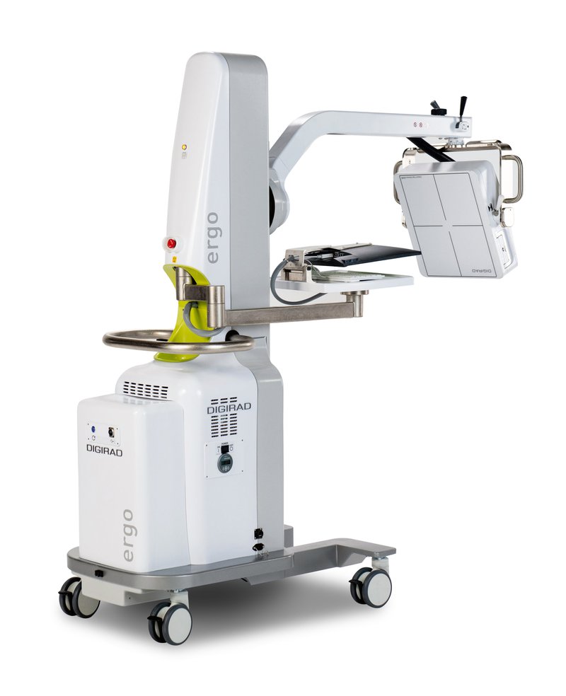
The Ergo revolutionizes the portable camera using solid-state flat panel detector technology. Since the Ergo does not use Photo-Multiplier tubes in its detector, it is quiet, compact, light, and easy to maneuver.
Imaging quality is high and will allow the physician to get real-time confirmation of whether the particulate dose and embolization have been successful. This means decisions can be made more quickly and use of clinical staff can be optimized.

Ergo features
Ergo has a large field of view detector. The system delivers an intrinsic spatial resolution of 3.25mm, energy resolution of 7.9% and count rate capabilities greater than 5 Mcps. It is lightweight and easy to move without the use of a motor, and at 29” wide, has a small footprint, easily fitting through doors.
Another plus for the Ergo gamma camera is that it’s very versatile. It seamlessly moves for use in areas such as general radiology, women’s health, pediatrics, surgery and trauma. This makes it a great choice not only for Y90 imaging, but for many purposes within the medical facility.
Six high performance collimator options (LEAP, LEHR, Pinhole, MEAP, Diverging, and MBI) provide the capability to provide outstanding imaging quality for a wide range of procedures for energy ranges from 50 to 350 keV. Quick release latch mechanisms and flip-up handles make changing collimators simple and fast. An optional collimator storage cart provides easy and convenient accessibility and storage for up to five collimators and one breast imaging accessory.
Point-of-care imaging is changing the way the radiology department operates for the better. It’s a great option for streamlining Y90 procedures, optimizing use of staff and space, and reducing risks associated with patient transport and longer times under anesthesia.
The in-suite imaging advantage
Yttrium-90 (Y90) studies form a key part of the treatment plan for many liver cancer patients, however the procedure is often lengthy and inefficient for both patients and clinics.
There is a better way to conduct Y90 studies, using mobile imaging. This removes back and forth between a treatment room and radiology, allowing medical facilities to optimize their use of staff and camera equipment.
The Ergo gamma camera is an ideal option for Y90 studies as well as many other imaging needs. Its portability and versatility make it a great addition to any clinical setting.


